Her lost arm gave its nerve to her leg, and her toe became a thumb
A 48-year-old woman survived a shark attack in New Caledonia that took her left hand, the thumb and two fingers of her right hand, and 16 centimetres of the main nerve of her leg. Over six months and seven major operations, her surgeons used what the attack had left to rebuild what it had taken. A year later she was walking, and dancing.
- Specialty
- Plastic and reconstructive microsurgery
- Patient
- Woman, 48
- Mechanism
- Shark attack, New Caledonia
- Hands
- Left hand lost at the forearm; right thumb, index and middle fingers lost
- Leg
- 36 × 24 cm wound of the lower back and left thigh; 16 cm of the sciatic nerve missing; open ankle fracture
- Reconstruction
- 6 months, 7 major operations
- Nerve
- Bridged with the ulnar nerve from her amputated arm, on its own blood supply
- Thumb
- Rebuilt from her left big toe
- At 12 months
- Walking with a stick, independent, dancing
- day 0the attack
- stage 1clearing the wounds
- stage 2the right hand
- stage 3leg and thumb
- 12 monthswalking
What the shark took
The report does not describe the attack itself, only its result. A 48-year-old woman was attacked by a shark in New Caledonia, in the South Pacific. She was stabilised there, and the surgeons diverted her bowel with a colostomy, before she was transferred to the authors’ plastic surgery teams in Sydney for the reconstruction.
The list of injuries was long. Her left hand had been amputated at the lower forearm. On her right hand, the thumb had been lost at the knuckle at its base and the index and middle fingers at their first bones. Across the back of her trunk and her left thigh there was a wound 36 by 24 centimetres. And in the depth of that wound, 16 centimetres of her sciatic nerve were simply missing. She also had an open fracture of her left ankle.
The sciatic nerve is the largest nerve in the body. It runs down the back of the thigh and controls the muscles that bend the knee and everything below it, as well as most of the feeling in the foot. Losing a long segment of it usually means a leg that cannot feel or move properly below the knee.
Shark bite injuries are among the most destructive in trauma surgery. The authors cite a review in which 62% of survivors needed an amputation or a disarticulation through a joint, and mortality approached 38%. Saving a limb so that it works again is rare.
Cleaning before building
The reconstruction was planned in stages, and over six months, from February to July 2023, she had seven major operations.
The first stage was preparation. In repeated operations the surgeons removed dead tissue and the contamination that sea water and a shark’s bite leave behind. The parts of her left forearm and right fingers that could not survive were revised into clean amputations. The huge wound on her back and thigh was covered with a dermal substitute, a synthetic layer that lets the body build a new bed of tissue before any skin or flap is placed on it.
A flap that could feel
Her right hand was her dominant hand, and it was the one she still had, so it came next. The surgeons covered its wounds with an anterolateral thigh flap: a piece of skin and fat from the front of the thigh, moved with its own artery, which they joined to the radial artery at the wrist.
They also made it sensate. The flap was taken with the small nerve that supplies feeling to that area of the thigh, the lateral femoral cutaneous nerve, and they joined it to a sensory nerve of the hand, the superficial radial nerve. Skin moved this way can learn to feel in its new place. A later operation refined the flap’s contour.
Using what the attack had left
The hardest problem was the 16-centimetre gap in the sciatic nerve. Ordinary nerve grafts, thin strips of a minor nerve taken from elsewhere, have no blood supply of their own when they are put in; they depend on the tissue around them to keep them alive. In a long gap, in a wound that had been contaminated and had a poor blood supply, that is a weak point. The alternative is a vascularised nerve graft, a nerve moved together with its own artery, so it stays alive from the moment it is placed.
And there was a nerve available that she no longer needed. The ulnar nerve runs down the inner side of the arm to the hand. Her left hand was gone, so the ulnar nerve in that arm had nothing left to serve. It was also thick enough to match the sciatic nerve and had a reliable artery of its own. The surgeons took a 16-centimetre length of it together with its artery, the superior ulnar collateral artery, on an 8-centimetre stalk.
To cover the back of her thigh they used a free flap of latissimus dorsi, the broad muscle of the back. They designed it as a “flow-through” flap: a branch of the flap’s own vessels, a serratus branch of the thoracodorsal system, was used to feed the ulnar nerve graft, while the flap’s main artery was joined to a large perforating vessel from the femoral artery. When they washed the nerve with saline, it bled briskly: it was alive.

The upper end of the graft was joined to the sciatic nerve’s stump, the lower end to its tibial division, the branch that runs on to supply the calf and the sole of the foot. The muscle flap was laid over it and covered with a thin skin graft.
Then the thumb. The thumb does about half the work of the hand, because almost every grip depends on it pressing against the fingers. The surgeons moved her left big toe to her right hand, on the first dorsal metatarsal artery, joining three nerves, one artery and two veins, repairing the tendons and fusing the knuckle at its base. Surgeons often slim down a big toe before turning it into a thumb. Here they chose not to: the toe came from her left foot, which had lost its feeling with the sciatic nerve, and trimming it would have removed tissue from the side of the new thumb that would matter most when she pinched.
PhotographHer right hand after the toe-to-thumb transfer, with fresh stitches. Tap to show.
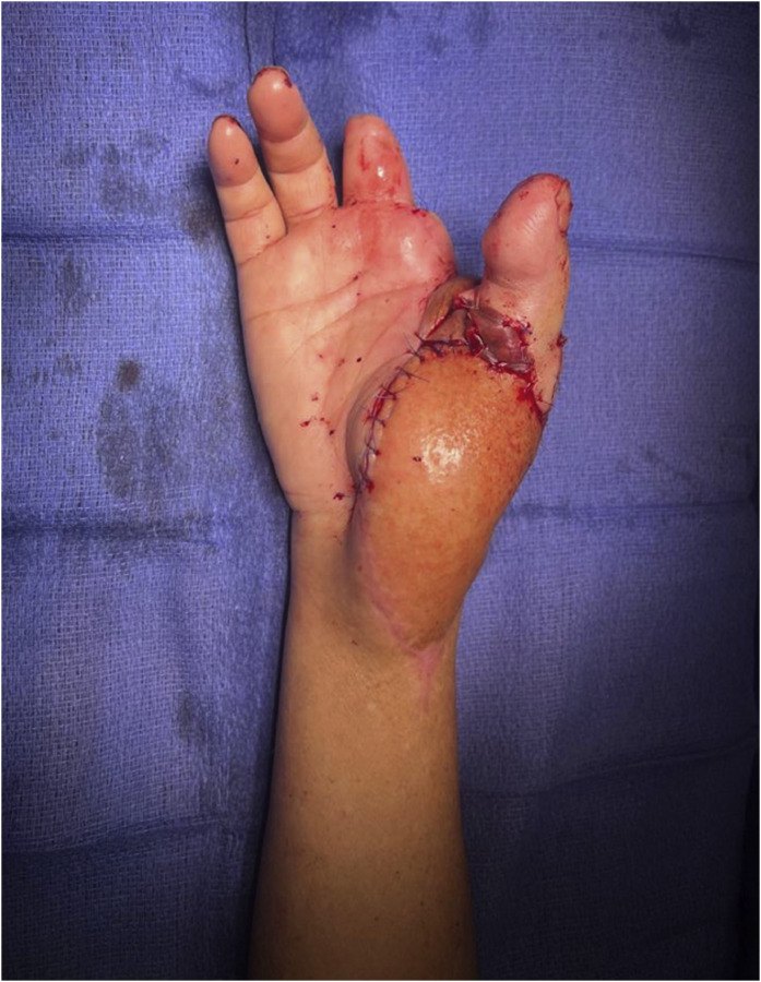
Two more procedures completed the stage. On the stump of her left forearm, the surgeons performed targeted muscle reinnervation: the cut ends of the nerves were redirected into nearby muscles, the median nerve into the flexor carpi radialis, the superficial radial nerve into the palmaris longus and the ulnar nerve’s upper stump into the triceps. A cut nerve with nowhere to go can form a neuroma, a painful knot of nerve fibres; giving it a muscle to grow into prevents that, and the reinnervated muscles can later produce signals to drive a powered prosthesis. And her left ankle was fused, so that the foot could not drop while the nerve recovered, giving her a stable platform to walk on.
A year later
Between the operations, rehabilitation focused on bearing weight, moving her joints and learning to use prostheses.
Twelve months after the nerve reconstruction, she was walking independently with a single stick. The muscles that bend her knee, supplied by the sciatic nerve, had recovered to a strength of 4 to 5 on the 5-point Medical Research Council scale. Feeling had returned in the sole of her foot, in the area supplied by the tibial nerve.
Her new thumb could oppose and pinch. She scored 7 out of 10 on the Kapandji test, which measures how far the thumb can reach across the hand, and her key pinch, the grip used to hold a key, reached 3.0 kilograms, about 55 to 60% of the other side. The flap on her hand had regained feeling, with two-point discrimination of about 12 to 15 millimetres. There was no sign of a painful neuroma in her forearm stump, and she had fine control of a myoelectric prosthesis, a powered artificial hand driven by her own muscle signals.
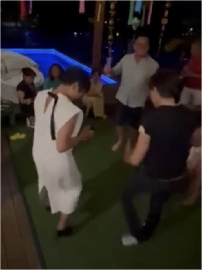
She was fully independent in her daily life, travelling, and dancing.
A nerve that brings its own blood supply
Vascularised nerve grafts are not new, but they are mostly used in the arm. The authors cite work by Doi and colleagues showing better survival of nerve fibres and better recovery with vascularised grafts than with conventional ones, and the situations where they are most useful: gaps longer than 6 centimetres, beds with a poor blood supply, wounds that also need a flap, injuries high up a nerve and nerves that have been without function for a long time. A 16-centimetre gap in a previously ischaemic, contaminated thigh met nearly all of these.
The authors say this is the first reported use of a vascularised ulnar nerve graft for a large segmental defect of the sciatic nerve, and the first report combining all of these techniques in one shark attack survivor. Vascularised sural nerve grafts, from the back of the calf, have been used for smaller sciatic injuries, but not for a gap this long.
They are also clear about the limits. This is a single patient, and with so many operations done together, no single one of them can be given credit for the recovery. Formal electrical testing of the nerve and muscles (EMG) was not done, because of the staged treatment, social and economic factors and the long distances she would have had to travel; the authors judged that her clear clinical recovery made such a test unlikely to change anything.
What this case teaches
A 48-year-old woman survived a shark attack that took her left hand, the thumb and two fingers of her right hand, and left a 36-by-24-centimetre wound across her back and thigh with 16 centimetres of her sciatic nerve gone. After the wounds were cleaned and prepared, her surgeons covered her right hand with a thigh flap whose nerve they joined so it could feel. They bridged the sciatic gap with the ulnar nerve from her amputated arm, carried on its own artery and fed through a flap of back muscle laid over it. They turned her left big toe into a right thumb, redirected the nerves in her forearm stump into muscle to prevent pain and drive a prosthesis, and fused her ankle. Seven major operations over six months later, she was walking with a stick, pinching with her new thumb, using a powered hand and dancing. In reconstruction after a catastrophic injury, sometimes the best spare parts are the ones the injury itself has freed up.
Was this case worth your time? Buy the next one a coffee ($3), or support MedicaseHub monthly.
Next case · MC-027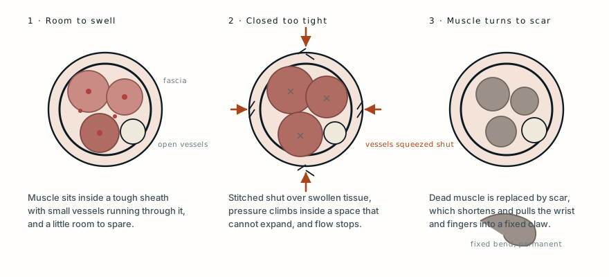 The shark did not cause the contractureThe bite took tissue. The hand was lost to a wound closed too tightly, and by the time she reached a second hospital it was already fixed.Read it →
Want the next case like this?
When a new case is written up, it comes to your inbox: the story, the diagram and what it teaches. No schedule and no filler. Unsubscribe in one click.
Free · no spam · your address is never shared · built with KitRead the original paper: doi.org/10.1016/j.jpra.2026.02.016
Adapted from Bishay J, Garrett H, Gillies R, Stewart D and Soliman B, “Multistage microsurgical reconstruction after catastrophic shark attack: Vascularized ulnar nerve grafting for a large segmental sciatic nerve defect,” JPRAS Open 2026;49:196–203, from Westmead Private Hospital and Royal North Shore Hospital, Sydney. The injuries, the stages and techniques of the reconstruction and the outcome measures are as reported there, summarised in the author’s own words; the figures on shark attack injuries and the evidence on vascularised nerve grafts are those the authors cite in their discussion, and the explanations of the anatomy and procedures are from standard references. The paper is published under the Creative Commons Attribution licence (CC BY 4.0), and the authors report written consent from the patient to publish her clinical details and images. The two photographs here are reproduced from Figures 3 and 4 under that licence, unchanged. The photographs of her wounds and of the operation are not reproduced. The diagram is original to MedicaseHub, is simplified, and may be reused freely under CC BY 4.0. This article is not medical advice. Read the full disclaimer.
Related cases
- The shark did not cause the contractureAnother shark bite, and a hand lost to what happened after it.
- They saved his ear by keeping it inside his bellyAnother reconstruction that borrowed tissue from somewhere unexpected.
- Two flaps, and no microscopeFlaps that kept their own blood supply, and why that matters.